SELECTED PROJECTS


Blastocyst Detection Cell-Net: Embryonic Cell Counting and Centroid Localization via Residual Incremental Atrous Pyramid and Progressive Upsampling Convolution
Reza Moradi Rad, Parvaneh Saeedi, Jason Au, and Jon Havelock
IEEE Access - PDF

In-vitro fertilization (IVF), as the most common fertility treatment, has never reached its maximum potentials. Systematic selection of embryos with the highest implementation potentials is a necessary step towards enhancing the effectiveness of IVF. Embryonic cell numbers and their developmental rate are believed to correlate with the embryo's implantation potentials. In this paper, we propose an automatic framework based on a deep convolutional neural network to take on the challenging task of automatic counting and centroid localization of embryonic cells (blastomeres) in microscopic human embryo images. In particular, the cell counting task is reformulated as an end-to-end regression problem that is based on a shape-aware Gaussian dot annotation to map the input image into an output density map. The proposed Cell-Net system incorporates two novel components, residual incremental Atrous pyramid and progressive up-sampling convolution. Residual incremental Atrous pyramid enables the network to extract rich global contextual information without raising the ‘grinding’ issue. Progressive up-sampling convolution gradually reconstructs a high-resolution feature map by taking into account short- and longrange dependencies. Experimental results confirm that the proposed framework is capable of predicting the cell-stage and detecting blastomeres in embryo images of 1 to 8 cell(s) by mean accuracies of 86.1% and 95.1%, respectively.
Blastocyst Detection Blast-Net: Semantic Segmentation of Human Blastocyst Components via Cascaded Atrous Pyramid and Dense Progressive Upsampling
Reza Moradi Rad, Parvaneh Saeedi, Jason Au, and Jon Havelock
IEEE International Conference on Image Processing (ICIP) - PDF

Components of a human blastocyst (day-5 embryo) and their morphological attributes highly correlate with the embryo's potentials for a viable pregnancy. Automatic semantic segmentation of human blastocyst components is a crucial step toward achieving objective quality assessment of such blastocyst. In this paper, a semantic segmentation system is proposed for human blastocyst components in microscopic images. The proposed Blast-Net features two novel components: a Cascaded Atrous Pyramid Pooling (CAPP) module to incorporate multi-scale global contextual priors, and a Dense Progressive Sub-pixel Upsampling (DPSU) module to recover the high-resolution prediction map. Experimental results confirm that the proposed method achieves the best-reported segmentation performance to date with a mean Jaccard Index of 82.85% for microscopic images of the human blastocyst.


Blastocyst Detection Predicting Human Embryos' Implantation Outcome from a Single Blastocyst Image
Reza Moradi Rad, Parvaneh Saeedi, Jason Au, and Jon Havelock
IEEE Engineering in Medicine and Biology (EMBC) - PDF

Only one-third of embryo transfer cycles via invitro fertilization, the most common fertility treatment, leads to a clinical pregnancy. Identifying embryos with the highest potentials for transfer is an essential step to optimize in-vitro fertilization outcome. However, human embryos are complicated by nature and some of their developmental aspects has still remained a mystery to expert biologists. In this paper, the firstever attempt is made to estimate probability of implantation using a single blastocyst image. First, a semantic segmentation system is proposed for human blastocyst components in microscopic images. Second, a multi-stream classification model is proposed for the prediction of embryos' implantation outcome. The proposed classification model features an architectural component, Compact-Contextualize-Calibrate (C3) to guide the feature extraction process and a slow-fusion strategy to learn cross-modality features. Experimental results confirm that the proposed method delivers the first-reported implantation outcome prediction via a single blastocyst image to date with a mean accuracy of 70.9%.


Blastocyst Detection Human Blastocyst's Zona Pellucida Segmentation via Boosting Ensemble of Complementary Learning
Reza Moradi Rad, Parvaneh Saeedi, Jason Au, and Jon Havelock
Informatics in Medicine Unlocked - PDF

Characteristics of Zona Pellucida (ZP), particularly its thickness, is a key indicator of human blastocyst (day-5 embryo) quality. Therefore, ZP segmentation is an important step towards achieving automatic embryo quality assessment. In this paper, a novel approach based on boosting ensemble of hybrid complementary learning is proposed to segment Zona Pellucida in human blastocyst images. First, an inner-ZP localization method is proposed to separate the ZP from the heavily textured area inside a blastocyst. Then, a deep Hierarchical Neural Network (HiNN) is proposed to segment ZP area. The hierarchical nature of the proposed network enables learning features with respect to their spatial location in the embryo. Finally, a Self-supervised Image-Specific Refinement (SISR) strategy is proposed as a complementary step to boost the performance. The proposed system is a hybrid approach in the sense that the HiNN learns the intra-correlation among images, while the SISR takes into account the inter-correlation within the query image. Experimental results confirm that the proposed method is capable of identifying ZP area with average Precision, Recall, Accuracy and Jaccard Index of 85.2%, 92.0%, 95.6% and 78.1%, respectively. The proposed HiNN system outperforms state of the art by 4.9% in Precision, 11.2% in Recall, 3.6% in Accuracy and 10.7% in Jaccard Index.


TE Segmentation A hybrid approach for multiple blastomeres identification in early human embryo images
Reza Moradi Rad, Parvaneh Saeedi, Jason Au, and Jon Havelock
Computers in Biology and Medicine - PDF

Analyzing the shape, size, and motion of the cells, as well as other time-related changes, facilitates embryo quality assessment. However, the ambitious 3D-like side-lit appearance of the embryo, occlusion, transparency of cells and artifacts such as fragmentation make automatic detection of blastomeres (embryonic cells) a challenging task. In this paper, an automated non-invasive approach is proposed to identify multiple blastomere cells inside an embryo at different growth stages. In particular, the proposed method aims to identify up to 8 blastomeres in microscopic human embryo images of days 1-3. The proposed system is a hybrid approach that aggregates both models and features capturing global and local characteristics to locate the boundaries of each blastomere. Experimental results on a large dataset confirm that the proposed method identifies blastomeres with average Precision, Recall, and Overall Quality of 85.9%, 85.3%, and 76.5%, respectively.


UES Unification of Data Embedding and Scrambling
Reza Moradi Rad, KokSheik Wong, and Jing-Ming Guo
IEEE Transactions on Image Processing - PDF

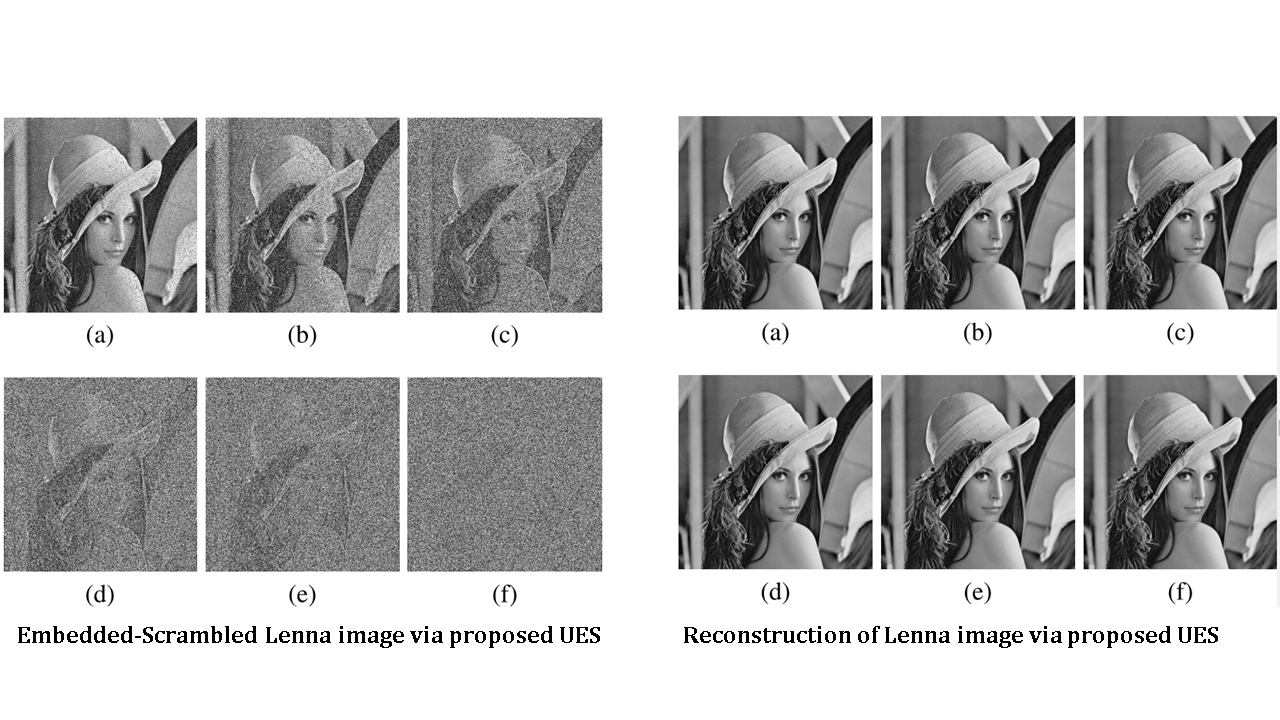
Conventionally, data embedding techniques aim at maintaining high-output image quality so that the difference between the original and the embedded images is imperceptible to the naked eye. In this paper, a unified data embedding-scrambling technique called UES is proposed to achieve two objectives simultaneously, namely, high payload and adaptive scalable quality degradation. Given a desirable quality (quantified in SSIM) for the output image, UES guides the embedding-scrambling algorithm to handle the exact number of pixels, i.e., the perceptual quality of the embedded-scrambled image can be controlled. In addition, the prediction errors are stored at a predetermined precision using the structure side information to perfectly reconstruct or approximate the original image. In particular, given a desirable SSIM value, the precision of the stored prediction errors can be adjusted to control the perceptual quality of the reconstructed image. Experimental results confirmed that UES is able to perfectly reconstruct or approximate the original image with SSIM value >0.99 after completely degrading its perceptual quality while embedding at 7.001bpp on average.


AGM Reversible Data Hiding by Adaptive Group Modification

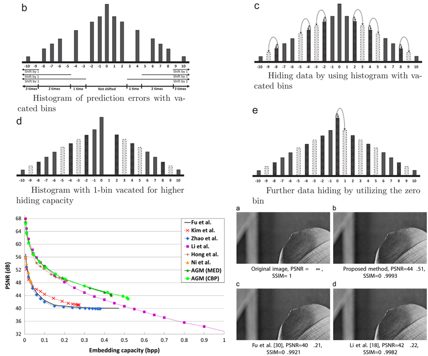
In this work, the conventional histogram shifting (HS) based reversible data hiding (RDH) methods are first analyzed and discussed. Then, a novel HS based RDH method is put forward by using the proposed Adaptive Group Modification (AGM) on the histogram of prediction errors. Specifically, in the proposed AGM method, multiple bins are vacated based on their magnitudes and frequencies of occurrences by employing an adaptive strategy. The design goals are to maximize hiding elements while minimizing shifting and modification elements to maintain image high quality by giving priority to the histogram bins utilized for hiding. Furthermore, instead of hiding only one bit at a time, the payload is decomposed into segments and each segment is hidden by modifying a triplet of prediction errors to suppress distortion. Experimental results show that the proposed AGM technique outperforms the current state-of-the-art HS based RDH methods. As a representative result, the proposed method achieves an improvement of 4.30 dB in terms of PSNR when 105,000 bits are hidden into the test Lenna image.


Image Forgery Digital Forgery Detection in Image

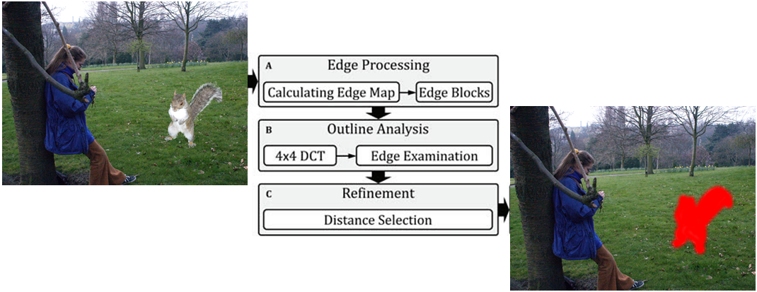
The advent of user-friendly yet powerful editing softwares has cast doubt on the authenticity of digital images. Therefore, developing reliable detection techniques is of great importance to verify the originality of a given image. In this work, a forgery detection technique based on the analysis of edge information is proposed. Unlike the conventional methods, the proposed technique is not restricted to the traces left by the act of double compression, but instead it allows the input image to be singly compressed or uncompressed. Experimental results confirmed that proposed method is able to localize forged area when the forged image is not double compressed.


Image spam Reversable Data Embedding in Scrambled JPEG Image

In this work, an efficient DCT sign prediction method is proposed. Unlike the conventional methods that depend on information from both spatial and frequency domains, the proposed method operates solely in the frequency domain by exploiting the pixel value patterns represented by the corresponding DCT basis vectors. In particular, each block is classified into five categories, namely, complex-pattern, complex-nonpattern, simple-smooth, simple-pattern and simple-texture, and each is treated differently using the proposed predictor. The proposed sign prediction method is then applied to realize reversible data embedding using sign information in a scrambled JPEG compressed image. This work is the first of its kind in using sign information for data embedding purposes. Basic performance of the proposed sign prediction and the proposed reversible data embedding method in scrambled JPEG image are verified using standard test images.


Image spam Image Quality Assessment

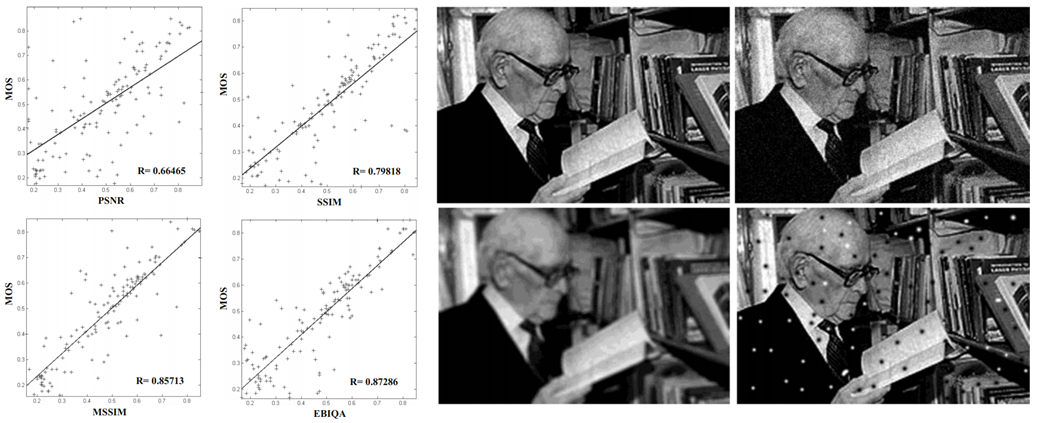
There are many applications for Image Quality Assessment (IQA) in digital image processing. Many techniques have been proposed to measure the quality of an image such as Peak Signal to Noise Ratio (PSNR), Structural Similarity (SSIM), and Mean Structural Similarity Index Measurement (MSSIM). In this paper, a new technique, namely, Edge Based Image Quality Assessments (EBIQA) is proposed. The proposed technique is based on different edge features which are extracted from original (distortion free) and distorted images. The new approach was implemented and tested using different images which have been taken from A57 and WIQ image databases. The experimental results show that the functionality of the EBIQA technique is better than the state of art IQA techniques. The proposed technique is consistent with the mean opinion score which makes it suitable for automatic image quality assessment.


Image spam Image Spam Detection

Many techniques have been proposed to combat the upsurge in image-based spam. All the proposed techniques have the same target, trying to avoid the image spam entering our inboxes. Image spammers avoid the filter by different tricks and each of them needs to be analyzed to determine what facility the filters need to have for overcoming the tricks and not allowing spammers to full our inbox. Different tricks give rise to different techniques. This work surveys image spam phenomena from all sides, containing definitions, image spam tricks, anti image spam techniques, data set, etc. We describe each image spamming trick separately, and by perusing the methods used by researchers to combat them, a classification is drawn in three groups: header-based, content-based, and text-based. Finally, we discus the data sets which researchers use in experimental evaluation of their articles to show the accuracy of their ideas.